Shielding Considerations and Requirements for a
PET/CT Scanner Facility
Ning Yue, Ph.D., DABR
Chief of Physics, Professor of Radiation Oncology
The Cancer Institute of
Positron Emission Tomography (PET)
is being used more and more not only as an imaging modality for diagnosis but
also for the purpose of delineation of target volume in radiation therapy. The physical principle of the PET
tomography is to coincidentally detect the two photons that are produced during
a positron-electron annihilation process. The photons such produced possess an
energy of 511 keV, which high level distinguishes a PET/CT scanner from
conventional CT scanner as far as radiation protection is concerned and has to
be taken into consideration in the shielding design of a PET/CT scanner
facility.
Unlike conventional CT scanner
x-ray sources, the radiotracers used in PET imaging have relatively short
half-lives and a constant dose rate cannot be assumed in the PET dose
calculations. Furthermore, since most of the annihilation incidents take place
inside patient body, a significant amount of radiation is absorbed by the
patient body itself. Therefore, in general, the radiation dose at a point of interest
over a time t should be computed as:
![]() (1)
(1)
ˇˇ
where L
is effective dose equivalent rate, A(0)
is the radiotracer strength at time 0,
d is the distance between the patient
and the point of interest, T1/2
is the half-life of the radionuclide, and ![]() is patient body attenuation factor.
is patient body attenuation factor.
In a typical PET/CT scanner
facility, in addition to PET/CT scanner room, there is usually a patient uptake
room where patient is kept in quiet and resting state for appropriate amount of
time (in the range of 30-90 minutes). The radiation dose contributions from
both the scanner room and the uptake room should be included, and equation 1
should then be expressed as:
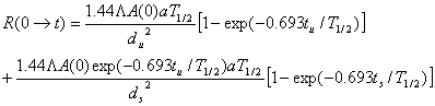 (2)
(2)
ˇˇ
ˇˇ
where tu
is uptake time, ts is
scanning time, and t= tu + ts, du
is the distance from the point of interest to the patient in the uptake room,
and ds is the distance
from the point of interest to the patient in imaging position. It is also
assumed that the contribution after the imaging is negligible.
The photons from the transmission
component of PET/CT scanner are usually absorbed by the detectors and patients,
and are considered to be negligible. The radiation contribution from the CT
component can be computed as for a conventional CT scanner. The rest of the
shielding calculations are similar to the conventional one except that the
attenuation coefficients of shielding materials are different for the PET
component and the CT component.
Though 18F is the most commonly used radionuclide in PET applications, different types of radionuclides have also been considered or used as radiotracers. Those radionuclides have different half-lives, which can be as short as 76 second (82Rb), and can be as long as 4.2 days (124I). Some of the radionuclides can even emit photons with energies much higher than 511 keV (e.g., 1693 keV photons emitted from 124I). Depending on the type of radionuclide to be used, these characteristics need to be taken into account in the PET facility shielding design.